 English
  Ελληνικά
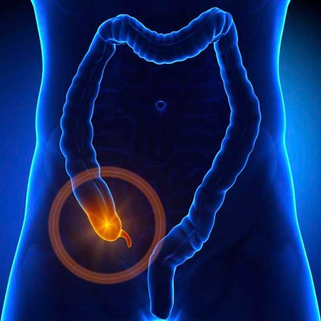
Acute appendicitis is an inflammation of the appendix that hangs from the cecum (the first part of the large intestine) and takes the form of an intestinal follicle.
It is due to the obstruction of its lumen by coprolith, hypertrophy of its follicles, seeds, parasites or tumors (carcinoid). The obstruction results in its dilation because the secretion of its mucosa (the inner wall) does not stop, while at the same time the multiplication of germs begins. When the cause of the obstruction ceases to exist the inflammation can subside. In most cases, however, the gradual dilation of the appendix affects the perfusion of the wall, where it can lead to perforation and peritonitis.
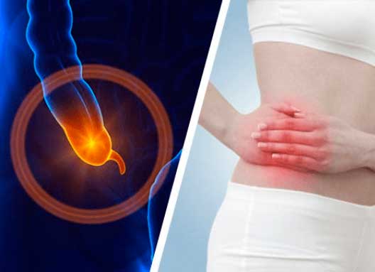
Acute appendicitis is the most common cause of abdominal pain in the second and third decades of life with a slight predominance in men over women.
Abdominal pain is the predominant symptom. It is initially located periomphalically and is diffuse and then shifts to the lower right quadrant of the abdomen (right iliac fossa) and becomes more intense and continuous. The pain is aggravated by pressure, coughing, walking or movement. The pain may be accompanied by anorexia, nausea or vomiting, fever (not very high), constipation or diarrhea.
The signs found on physical examination have to do with the location of the appendix and the progression of inflammation and are:
- Tachycardia and temperature raise
- The patient takes the position that comforts him, on the right side or on the back with the legs folded
- Mac Burney point: Sensitivity and contraction when palpated and pressed between the middle and outer third of the line joining the navel to the right anterior upper iliac spine
- Blumberg point or reflux: The patient complains of more intense pain when the abdominal wall pressure rises abruptly and is due to an extension of inflammation in the peritoneum
- Rovsing point: It is positive when left ventricular pressure or plexis causes right lower abdomen pain
- Thyroid point: When we rotate the right thigh that is bent inwards, it causes pain from contraction of the internal thyroid muscle if the appendix is in a pelvic position
- Psoas point: When the right leg is stretched, pain in the right groin manifests
- In the dactylic examination from the rectum or the vagina, the douglasious area is checked, which can be painful
From the laboratory test in the general blood test a slight increase of white blood cells with granulocytic type is observed. There is also usually an increase in CRP. If the appendix is located near the ureter then there may be pyocytes or red blood cells in the urine due to inflammation.
Ultrasonogram and CAT scan are useful tests for diagnosis because in addition to diagnosing the disease, they help to rule out other diseases in the area that cause similar pain.
The treatment of acute appendicitis is appendectomy which can be performed by laparotomy or laparoscopy. The indication for appendectomy should be given in time before its perforation. Laparoscopy is a method of choice of differential diagnostic problems in patients with right iliac fossa pain, especially in women, because it provides the possibility of complete control of the intra-abdominal organs.
In case of rupture of the appendix, in addition to appendectomy, peritonitis should be treated with rinsing of the venter, drainage placement and intravenous administration of antibiotics. If the rupture is vallated by the body (major omentum and intestinal helices) then plastron is created. This condition is treated conservatively, because appendectomy in this highly inflammatory local condition is sometimes not possible and then it is recommended to be performed three months later.