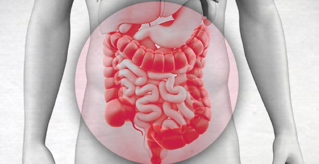

The peritoneum is a serous membrane composed of thin layers of loose connective tissue and is covered by a line of mesothelial cells, originates from the mesoderm and begins to form during the 4th week of fetal life.
It rubs the space between the organs of the venter (visceral peritoneum) and the abdominal wall (parietal peritoneum). The parietal peritoneum covers the posterior surface of the anterior abdominal wall, the lateral abdominal wall, the inferior surface of the diaphragm, the anterior surface of the retroperitoneal viscera and the pelvis. The visceral peritoneum covers the intestine, the intra-abdominal viscera and the mesentery.
Its function is the production of peritoneal fluid which minimizes friction and facilitates the free movement of the intra-abdominal viscera. It is also an elective barrier to the movement of fluids, soluble substances and foreign bodies and reduces inflammation. Finally, it contributes to the support of the intra-abdominal organs, which is achieved through the creation of ligaments.
The absorptive function of the peritoneal membrane is enhanced by structures such as the ora, which communicate with the subjacent afferentia, the septal peritoneum and the white spots on the omentum. The parietal peritoneum does not have such structures.
WHAT IS PERITONEAL CARCINOMATOSIS
Intra-abdominal cancer is dispersed in three ways:
- Through the portal and systemic venous circulation cells migrate and settle in other organs.
- Through the afferentia, cancer cells migrate to lymph nodes.
- Through erosion of the organ wall free cancer cells disperse into the venter.
Cells that disperse into the venter can survive and grow, when conditions allow, in multiple intra-abdominal implantations. This condition is called peritoneal carcinomatosis or peritoneal malignancy.
There are some types of cancer that are more common in peritoneal carcinomatosis. Such are the primary tumors of the peritoneum, such as mesothelioma, appendicitis cancer (pseudomyxoma), ovarian cancer and colon cancer. But potentially any cancer of the intra-abdominal and other organs can lead to peritoneal carcinogenesis.
This condition is still considered by many surgeons and oncologists as the final stage of an incurable disease. But the systematic clinical and laboratory research of Paul Sugarbaker, his colleagues and other researchers has shown spectacular results and increased survival or even cure in many cases.
Hyperthermic endoperitoneal chemotherapy in combination with cytoreductive surgery is the treatment of choice in which tumors that have developed inside the venter are surgically removed and then the application of hyperthermic chemotherapy leads to the destruction of the residual disease that may exist after the surgical resection of visible lesions.
The HIPEC method is based on the fact that there is an anatomical barrier (peritoneal-plasma barrier) between the peritoneum and plasma which prevents the rapid absorption of high molecular weight chemical compounds administered to the venter. Thus cytostatic drugs, most of which are high molecular weight compounds, have the property of settling on the peritoneal surfaces and exerting prolonged and intense their pharmacological properties, while at the same time being slowly absorbed into the systemic circulation. The penetration depth of the drugs is limited and it is not possible to destroy implantations with a maximum diameter greater than 2-3mm.
WHAT IS CYTOREDUCTIVE SURGERY
The purpose of cytoreductive surgery is the maximum reduction or even complete extinction of the disease from the venter. This is done with peritoneal resections, as described by P.H. Suarbaker and they are:
- Greater omentum and spleen excision
- The right and left subdiaphragmatic peritonectomy
- Pelvic peritonectomy
- Cholecystectomy and omental bursa excision
- The lesser omentum excision
- Right and left side-wall peritonectomy.
These excisions are performed with the help of diathermy, which has a 2mm ball at its end, which serves to separate the normal tissues from the cancer load.
HOW THE HYPERTHERMIC ENDOPERITONEAL CHEMOTHERAPY IS PERFORMED (HIPEC)
HIPEC is performed with the help of a special apparatus, consisting of a central unit, in which the chemotherapeutic and the solution are heated, one with two pumps that help the introduction and return of the solution and a system of tubes, two of which introduce the drugs into the venter and the other two return them to the central unit.
The solution of the chemotherapeutic agents with their solvent is subjected to the effect of high temperature and enters the venter at 44oC and is maintained at a temperature of 42.5-43oC. At this temperature it is possible to destroy cancer implantations without destroying the DNA of normal cells.
HIPEC techniques are two: the open and the closed method.
In the open method, the surgeon applies the Coliseum technique, i.e. he splices the skin on a special hook located around the abdomen to create a large single cavity and with his hands he makes sure that the drug is diffused to all tissues.
In the closed method, four drainages are placed, usually two in the hemidiaphragms and two in the pelvis and it is done at the end of the surgery after the abdomen is closed.
ASSESSMENT OF THE EXTENT OF PERITONAL CARCINOMATOSIS
The extent and distribution of peritoneal malignancy is one of the most important prognostic factors of peritoneal malignancy. It can be assessed either intra-operatively after the dehiscence of the venter or laparoscopically. Preoperative CAT scan, MRI scan and PET (positron emission scan) are important in assessing the extent of the disease. Staging and scoring of peritoneal carcinomatosis are useful in identifying suitable patients who will benefit from cytoreductive surgery and hyperthermic endoperitoneal chemotherapy (HIPEC), avoiding unnecessary aggressive treatments. We can also compare the results of studies from different centers and finally we can have a more accurate prognosis of the outcome of the therapeutic intervention.
To date, different methods have been used to assess the extent of peritoneal malignancy and the Peritoneal Cancer Index (PCI) has prevailed.
To define the PCI, the venter is divided into 13 anatomical sections and the magnitude of the lesion is taken into account. The anatomical sections are defined as AR and the magnitude of the implantations as LS (FIGURE 1).
The AR-0 section includes the medisection, the transverse colon and the major omentum. AR-1 includes the upper surface of the right lobe of the liver, the lower surface of the right hemidiaphragm and the Morisson's pocket. The AR-2 area includes the epigastric fat, the upper surface of the left lobe of the liver the lesser omentum and the falciform ligament. The AR-3 section includes the lower surface of the left hemidiaphragm, the anterior and posterior surface of the stomach, the tail of the pancreas, and the spleen. The AR-4 section includes the descending colon and the left parietal groove. AR-5 includes the sigmoid and lateral pelvic peritoneum. AR-6 includes the female's internal genitals, the posterior surface of the bladder, the Douglas space, and the rectosigmoid. AR-7 includes the base of the caecum, the appendix, and the right lateral pelvic peritoneum. AR-8 includes the ascending colon and right paracolic groove. AR-9 is defined as the upper jejunum, AR-10 as the inferior jejunum, AR-11 as the upper ileum and AR-12 as the inferior ileum.
The largest implantation size is taken into account to determine the criteria for the size of the tumor. The criteria for estimating the size of the implantations are as follows: the volume is judged as LS-0 when there is no macroscopically visible tumor, as LS-1 when the diameter of the implantation is less than 5mm, as LS-2 when the diameter is between 5mm- 5cm and as LS-3 when the diameter is greater than 5 cm or confluent implantations of various sizes are discovered.
The peritoneal cancer index is obtained by summing the size of the implantations for all 13 sections.
PCI is of prognostic importance in ovarian cancer, colorectal cancer, peritoneal sarcomatosis and the aggressive form of peritoneal mesothelioma. In peritoneal pseudomyxoma and low malignancy mesothelioma it has no prognostic significance, because perfect cytoreduction is possible, even when the index is 39, in which case there is complete occupation of the peritoneal surfaces.
DEGREE OF MALIGNANCY - BIOLOGICAL BEHAVIOR OF THE TUMOR
The degree of malignancy of the tumor, the histological type and the biological behavior of the tumor play an important role in both the prognosis and the treatment planning.
Peritoneal dispersion in relation to hematogenous and lymphogenous requires less complex biological processes. The emergence of peritoneal carcinomatosis is related to the histological type, the degree of malignancy of the primary tumor, as well as the presence or absence of ascites.
Medium or high malignancy cancers cause accidental dispersion and implantations in the venter, in areas close to the primary focus. On the surface of cancer cells there are adhesion molecules that facilitate implantation in the peritoneal surfaces. Presence of ascites facilitates the transport of cancer cells to more distant locations within the venter. Also, tumors that produce mucus, even of low malignancy, can cause extensive and massive dispersion to all surfaces of the venter, as occurs in tumors of the appendix and ovaries. This is because cancer cells have a low adhesion capacity, so they do not adhere directly to the peritoneal surfaces and at the same time mucus production increases the amount of fluid circulating in the venter and the transported cancer emboli can be implanted anywhere in the venter.
Dispersion can be the result of spontaneous tumor rupture or iatrogenically during surgery.
ADEQUACY OF CYTOREDUCTIVE SURGERY
The adequacy of cytoreductive surgery is the most important factor in the prognosis of the disease and is always assessed after the end of surgery. Thus, four types of surgeries have been identified:
CC-0 operations are those in which there is no macroscopically visible tumor after the operation. CC-1 operations indicate that the residual tumor has a maximum diameter of less than 2.5 mm. In CC-2 operations the residual tumor has a maximum diameter greater than 2.5 mm and less than 2.5 cm, while in CC-3 operations there is a residual tumor greater than 2.5 cm or confluent implantations of various sizes in different positions of the venter.
CC-0 and CC-1 type operations are considered completely cytoreductive when referring to low malignancy tumors. For tumors of high malignancy only CC-0 type operations are considered completely cytoreductive.
CC-2 and CC-3 surgeries are considered incomplete cytoreductive surgeries. The ability to perform complete cytoreduction depends on many factors, such as the degree of tumor malignancy, the extent and distribution of peritoneal malignancy and surgical skill.
OTHER FACTORS AFFECTING THE PROGNOSIS OF CYTOREDUCTIVE SURGERY
In cases of distant and non-resectable metastases, even if the surgeon manages to achieve complete cytoreduction, the survival is about the same as that of non-surgical patients. Of equal importance is the finding of metastases in groups of lymph nodes that are far from the primary focus.
Another prognostic factor is the general condition of the patient. Cytoreductive surgery with hyperthermic endoperitoneal chemotherapy is long-lasting, strains patients and is accompanied by high mortality. High-risk patients, such as patients with severe respiratory, heart, liver failure, severe eating disorders and intestinal obstruction are not recommended.
 English
  Ελληνικά