 English
  Ελληνικά
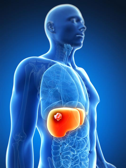
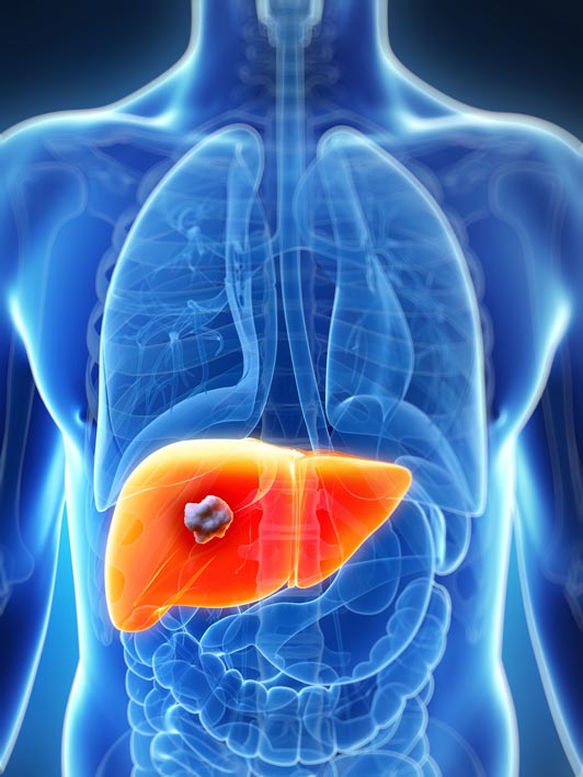
Liver cancer is a tumor that first appears in the liver tissue. There are different types, depending on the type of cancer cells.
Hepatocellular carcinoma (90%) is the most common type and starts from hepatocytes, liver cells.
A rare type is fibrolumellar carcinoma. It is usually well-defined in relation to hepatocellular carcinoma, which develops more invasively. It is treated like hepatocellular carcinoma.
Other types of cancer are tumors that grow in the liver but come from other organs, such as the large intestine, stomach or ovaries, and are called liver metastases.
ETIOLOGY
The main cause is hepatocirrhosis. In cirrhosis the normal liver tissue is gradually replaced by fibrous tissue and the hepatocytes do not grow and function normally.
- Risk factors that cause cirrhosis are:
- Chronic hepatitis B or hepatitis C virus infection
- Long-term alcohol abuse
- Hereditary liver diseases such as hemochromatosis and α1 antitrypsin deficiency
- Non-alcoholic fatty liver disease and non-alcoholic steatohepatitis
There are other less common conditions that increase the risk of cancer, such as autoimmune hepatitis, sclerosing cholangitis and Wilson's disease.
Also exposure to toxic agents (anabolic steroids, contraceptives) and ingestion of foods contaminated with aflatoxin (produced by a fungus that grows on nuts and rice).
epatitis A and C virus infection can cause cancer without preceding hepatocirrhosis.
CLINICAL PICTURE
Liver cancer usually has no symptoms in the early stages and the most common way it manifests is to find a mass on a random imaging test done for another reason.
Symptoms usually appear when the cancer has progressed and grown in size and are: pain on the right hypochondrium, weight loss, anorexia, malaise and jaundice.
DIAGNOSIS
The main method of diagnosis is the ultrasonogram, especially when combined with newer techniques (doppler, real time us).
Other diagnostic imaging techniques, such as various forms of CT and MRI scans and PET scans, are useful in both diagnosing and defining the surgical plan. MRI has surpassed CT in terms of sensitivity and ability to characterize a liver tumor.
Percutaneous liver biopsy offers little to the final diagnosis when all other tests are indicative. However, it is very useful in cases where the diagnosis is doubtful and in those where there is also hepatocirrhosis.
Blood tests include liver function tests (showing how well the liver is working), hepatitis tests, which show if the patient has or has had hepatitis B or C in the past and finally cancer indicators, which are indicative of the presence of liver cancer.
The most important cancer indicator is α-fetal globin (AFP) which is increased in 50-70% of cases of primary liver cancer.
Diagnostic laparoscopy helps detect peritoneal or extrahepatic spread, to determine if the healthy part of the liver is cirrhotic and to obtain biopsies under direct vision.
TREATMENT
The treatment approach depends on the stage of the disease (ie its extent), the condition of the liver (how well it works), as most cancers develop in hepatocirrhosis, the biological age (not the actual but the quality of health for the given age) and the general condition of the patient.
Treatment planning requires the involvement of an interdisciplinary oncology team of health professionals called an oncology board.
Due to the nature of the disease there is no single treatment. However, surgical resection of the tumor is a common component of all protocols in the treatment of hepatocellular carcinoma.
Surgical operations are the excision of the tumor with removal of a part of the liver called a partial hepatectomy and the hepatectomy with a liver transplant. Unfortunately many hepatocellular carcinoma patients may not be eligible for hepatectomy because tumor size, infiltration or thrombosis of major blood vessels do not allow it or due to insufficient function of the remaining liver due to cirrhosis.
The second type of treatment is the local destruction of the tumor by chemical or physical means. Such treatments include radiofrequency ablation (RFA), microwave (HIFU) or cryotherapy, and percutaneous alcohol or acetic acid injection.
Conservative methods of treatment of the disease are chemoembolization by catheterization of the hepatic artery (TACE), inoculation using small beads enriched with doxorubicin (chemotherapeutic) and radioembolization with microspheres of Iodine 131 or Yttrium 90.
Systemic chemotherapy responds to only about 25% of patients with non-surgical hepatocellular carcinoma. Various chemotherapeutic regimens have been used, such as XELOX (a combination of capecitabine and oxyplatin) and GEMOX (a combination of gemcitabine and oxaliplatin) that stop or slow the growth of the tumor. More modern drugs are sorafenib (Nexavar) which is a kinase inhibitor like lenbatinib (Lenvima).
Radiation therapy can be used in the case of a large tumor with few satellites (smaller tumors around it) and a sufficient portion of a healthy liver to save.