 English
  Ελληνικά
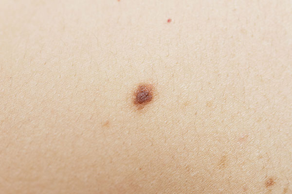
Nevi (commonly olives) are the clinical manifestation of proliferation and summation of cells normally found in the skin (melanocytes). However, there are other types of nevi that are not related to melanocytes such as blue nevus, phagesoral nevus and vascular nevus.
Melanocyte nevi are very common and can be found anywhere on the body but are more common in areas exposed to sunlight. They take various clinical forms and appear flat, round, slightly affected and pedicular.
Melanocyte nevi are distinguished in congenital and acquired.
Congenital nevi are present at birth and depending on the size are divided into small, medium and large (giant) when they exceed 20 cm. Giant pigmented nevi are relatively dangerous to develop melanoma and should be surgically removed.
Acquired nevi develop progressively throughout life with an increased frequency after adolescence, reach their maximum number around the age of 40 and then minimize the likelihood of their emergence.
In addition to the aesthetic problem they can cause, nevi are very important because they can develop into malignant melanoma which is the most dangerous skin cancer. Melanoma, if diagnosed early and radically removed, is effectively curable, but if it progresses profoundly or metastasizes, it has a bad prognosis.
To assess the degree of risk of a nevus, we consider the following:
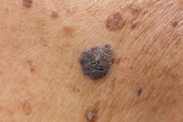
Α. Assymetry. Nevi where half the lesion is different from the other half are potentially more dangerous.
Β. Borders. Nevi whose borders are not uniform but aperiodic, lacy or serrated need testing.
C. Color. Nevi that do not have a uniform color distribution but different colors coexist (pink, brown, blue, black) need monitoring.
D. Diameter. Nevi larger than 6mm in diameter are more likely to become malignant.
Ε. Evolution. The change of ABCD in a short period of time (months to year) should mobilize us.
Also if the nevus shows itching, bleeding or emission of fluid should make us suspicious.
Surgical removal of a nevus is performed either for aesthetic reasons or for the risk of malignancy. The resection must be complete and with sufficient borders and the removed nevus must always be sent for biopsy.